Figure 1: Click on image to enlarge.
IMAGE OF THE WEEK
WEEK 39
HEPATIC LACERATION
The role of radiology in blunt liver trauma
|
|
|
|
|
Figure 1: Click on image to enlarge. |
|
|
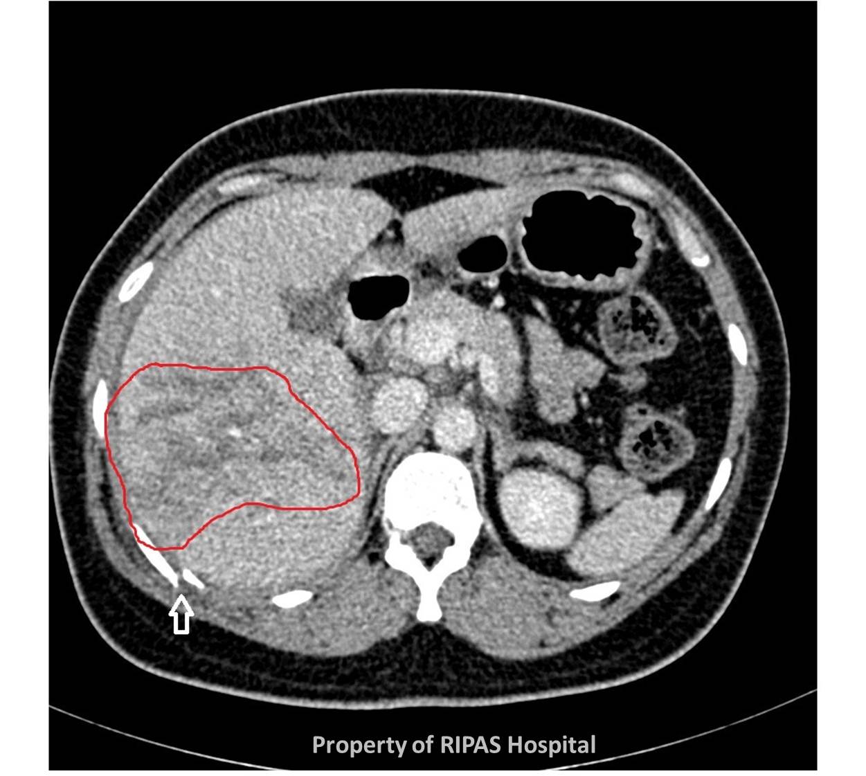
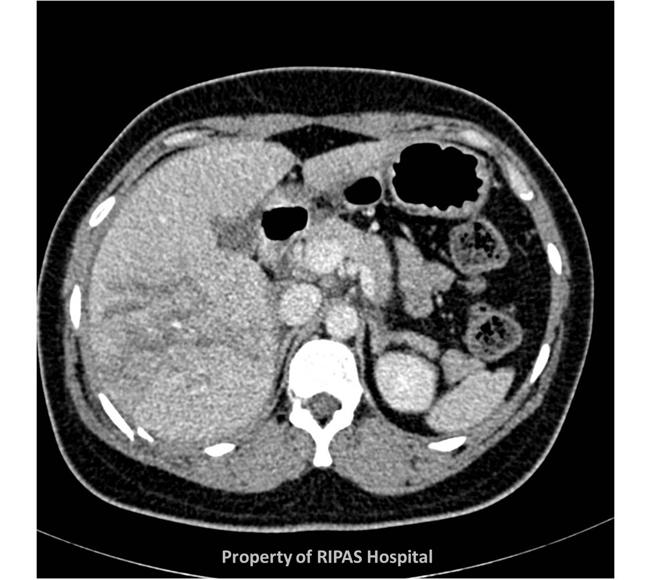
CT is the diagnostic modality of choice for the evaluation of blunt liver trauma in haemodynamically stable patients. It can accurately identify hepatic parenchymal injuries, quantify the degree of haemoperitoneum (presence of blood in the peritoneal cavity), and reveal associated injuries in other abdominal organs, retroperitoneal structures, and the gastrointestinal tract. Ultrasound may give a false negative result.
Hepatic lacerations are the most common type of parenchymal liver injury. They appear as irregular linear or branching low-attenuation areas on contrast-enhanced CT (within the red margin below). Note the adjacent rib fracture (white arrow).
|
|
|
Figure 2: Click on image to enlarge. |
Lacerations can be classified as superficial (≤3 cm in depth) or deep (>3 cm).
An in depth CT classification for liver trauma is also established which aids surgical decision making (see below).
Depending on the site of lacerations, likely complications to surrounding areas may be predicted. Lacerations that extend to the posterosuperior region of segment VII, the bare area of the liver, may be associated with retroperitoneal haematomas around the inferior vena cava and accompanied by adrenal hematoma. Lacerations that extend to the porta hepatis are commonly associated with bile duct injury and are thus likely to lead to the development of a biloma (an encapsulated collection of bile within the abdomen which occurs if there is bile duct disruption.
Images contributed and prepared by
Dr Ian Bickle, Consultant Radiologist, RIPAS Hospital & Diyana Mohamed, Trainee Intern/ Final year medical student ,University of Otago
All images are copyrighted and property of RIPAS Hospital.
![]()