
Figure 1a: CTPA showing a filling defect in the pulmary arteries on both sides as annotated in Figure 1b.
(Click on image to enlarge)
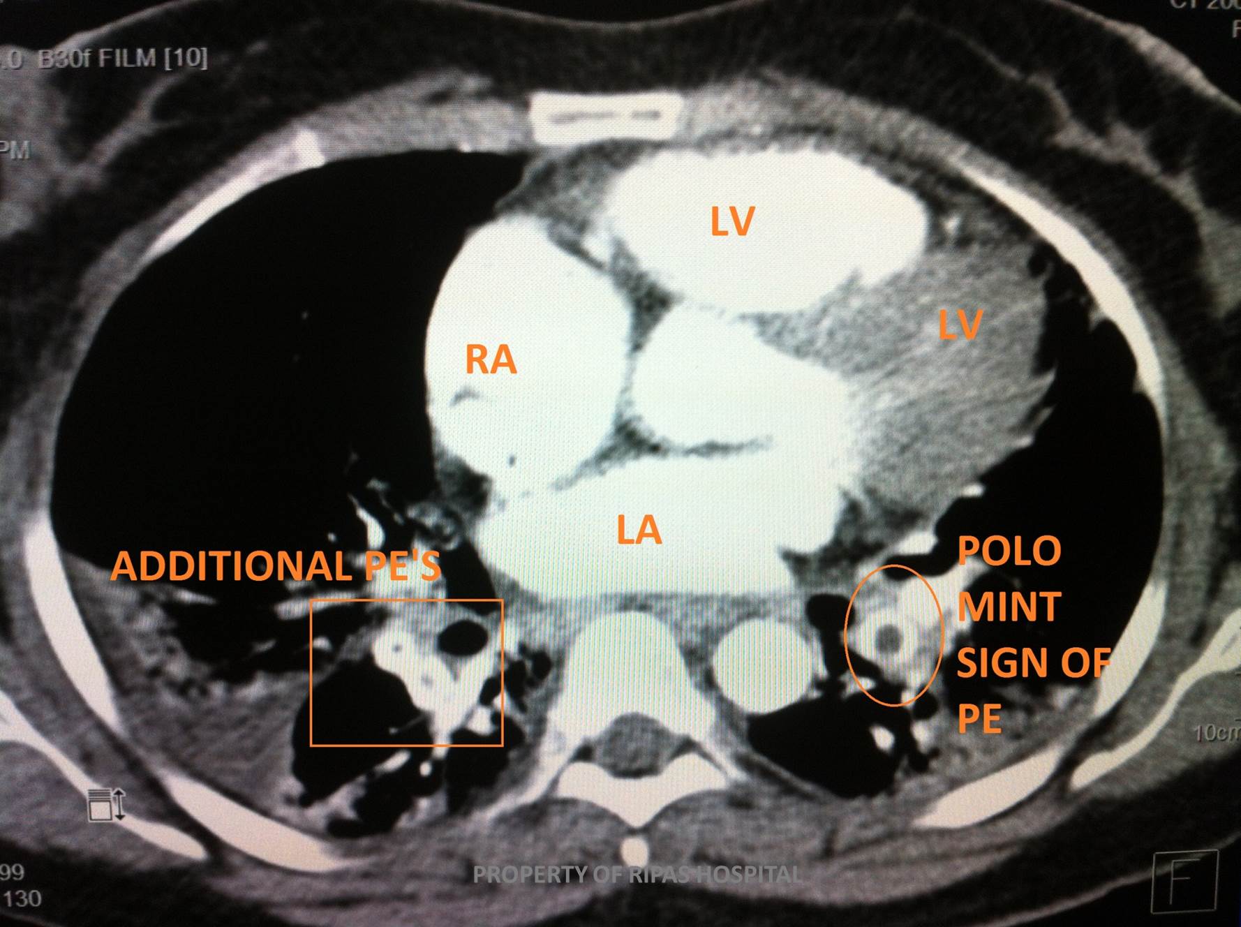
Figure 1b: Annotated CTPA of Figure 1a.
(Click on image to enlarge)
IMAGE OF THE WEEK 2013
WEEK 15
PULMONARY EMBOLUS
|
|
|
|
|
Figure 1a: CTPA showing a filling defect in the pulmary arteries on both sides as annotated in Figure 1b. (Click on image to enlarge) |
Figure 1b: Annotated CTPA of Figure 1a. (Click on image to enlarge) |
|
Pulmonary embolism is a common pathology and is now imaged almost exclusively with a CTPA (CT pulmonary angiogram).
With the use of a pump injector high speeds of intravenous contrast can be injected through a peripheral vein, with bolus track software on the CT management console, allowing image triggering when contrast illustrates the pulmonary venous system.
The concept is straightforward. Fill the pulmonary vasculature with dense iodinated contrast, and if an embolus is present it will show as a filling defect within (Figure 1). Multi-slice scanners enable excellent visualisation down to the sub-segmental vessels.
Emboli may be single or multiple (Figure 2). Large volume embolic disease and central emboli, such as the so called ‘saddle embolus’, may result in right heart strain. This may be identified on ECHO or on the CTPA itself as straightening of the intra-ventricular septum.
|
|
 |
|
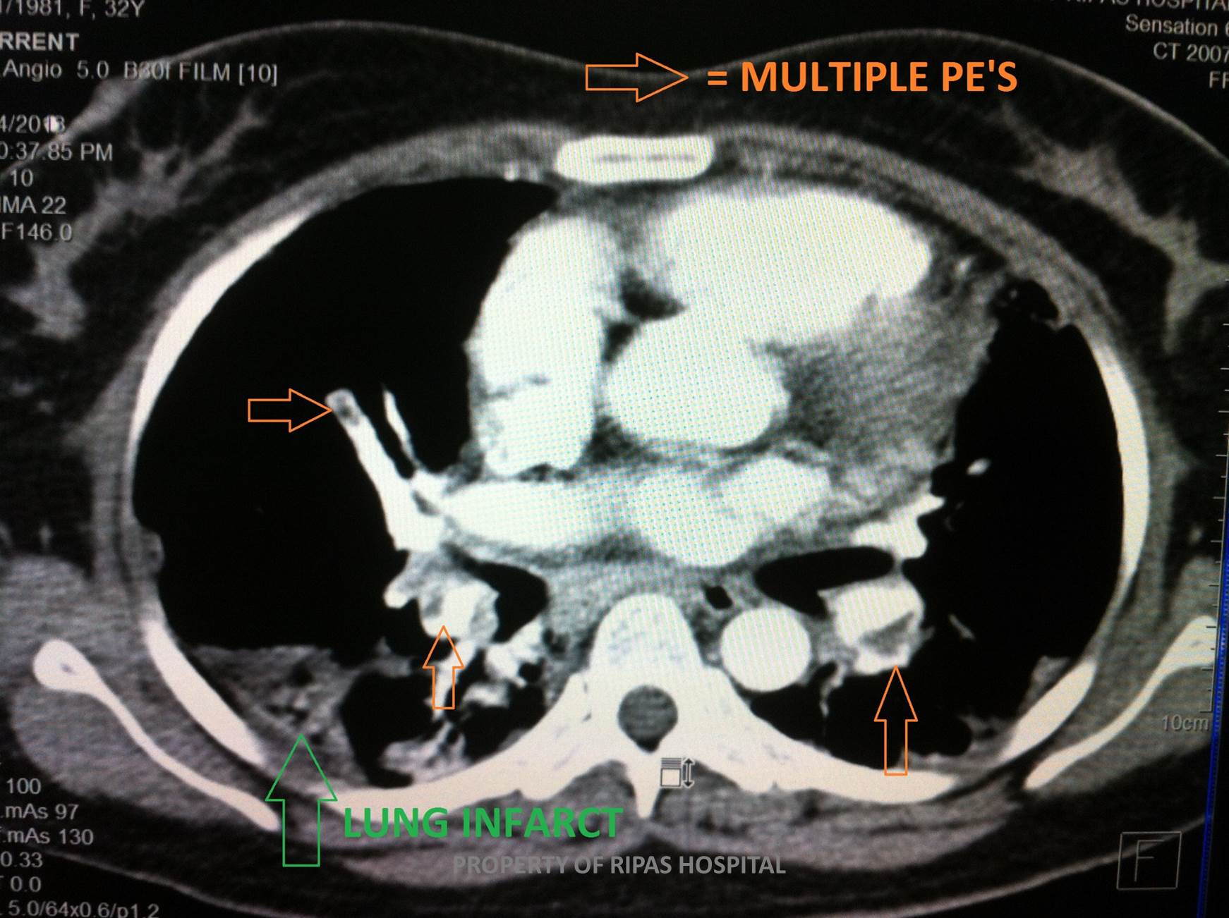
Figure 2a: CTPA showing multiple filling defects on both sides of the pulmonary arteries as annotated in Figure 2b. (Click on image to enlarge) |
Figure 2b: Annotated CTPA of Figure 2a showing multiple PEs. (Click on image to enlarge) |
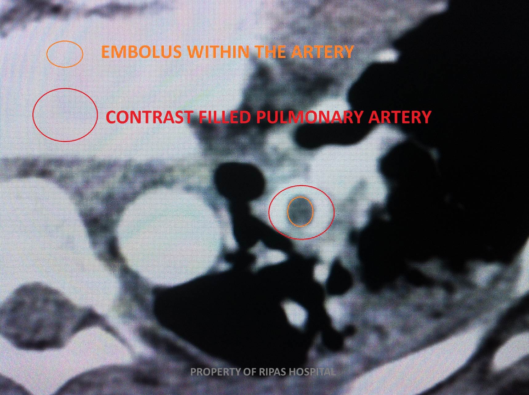
When an embolus is observed in the axial plane of a vessel (the vessel representing a circle) a rim of contrast may remain around the embolus – the ‘polo mint’ sign (Figure 3).
In a small proportion of cases, the distal lung may infarct – a triangular shaped area of consolidation is evident (Figure 2).
|
|
 |
|
Figure 3a: CTPA showing axial plane of a pulmonary artery with filling defect in the centre typical of the 'polo mint' sign. (Click on image to enlarge) |
Figure 3b: Annotated CTPA showing the 'polo mint' sign in an axial plane of a pulmonary artery. (Click on image to enlarge) |
Images and text contributed by
Dr Ian Bickle, Department of Radiology,RIPAS Hospital
All images are copyrighted and property of RIPAS Hospital.
![]()

